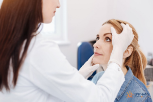
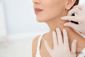
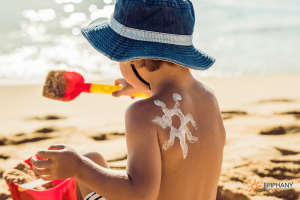
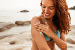
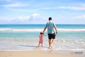
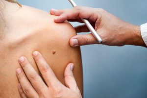
• Skip to primary navigation
  • Skip to main content
  • Skip to footer
COVID-19 Update  Teledermatology Appointments Now Available!
Skip to content

Epiphany Dermatology Logo

Header Right

  • About Us
    • Mission
    • Physicians
    • Extenders
    • Leadership
  • Services
  • Join
    • Overview
    • Partnering
    • Physician Jobs
    • Non-Physician Jobs
  • Locations
  • Appointments
  • Resources
    • Pay My Bill
    • Patient Forms
    • FAQs
    • Research
    • Blog
  • Locations
  • Book Appt

Considering Mohs Surgery on Your Face? 6 Important Facts About Facial Reconstruction After Mohs Surgery

If you’re considering Mohs surgery on your face, effectively treating your skin cancer is the main concern. However, you likely also have cosmetic concerns. While our primary goal as Mohs surgeons is to eliminate the cancer entirely, we also take reconstruction very seriously for the best aesthetic and functional results. We want to you to be cancer-free and develop a reconstructive approach that works best for you. If you’re considering Mohs surgery on your face, here’s what you need to know about Mohs reconstruction.

Does Every Patient Need Reconstruction After Mohs Surgery on the Face?

Not every patient needs reconstruction after their surgery, but for many, it’s the best choice. We opt for reconstructive surgery based on patient preferences and the specific anatomic location. We consider the type of closure needed, the local movement of the tissues, the crucial tissue functions, and how scar placement will affect long term aesthetic and functional outcomes. In my experience, patients heal faster (and often better) with reconstruction than they do without it. For example, when Mohs surgery is done on the rim of the nose, the eyelid, or the ear, we usually opt for reconstructive surgery. Each of these areas have a “free margin,” meaning it’s near the edge of the skin. If the defect heals without reconstruction, it can contract when the scar forms and distort the normal appearance of the area. A wide range of reconstructive options may be used including small side-to-side linear repairs, flaps, grafts, and various combinations of these.

When Does a Patient Decide If They Want Facial Reconstruction?

Choosing reconstruction after Mohs surgery on the face is a delicate balance between option and necessity. A good doctor will discuss your options with you up front and let you know what to expect based on his/her experiences. For example, when tumors are removed on the rim of the nose, defects will notch the nose if it is not reconstructed. So, I discuss potential reconstructive options with patients needing Mohs surgery on the nose from the beginning of the process. The doctor cannot determine where a tumor appears on a patient or how widespread it is prior to surgery, but they can be very upfront about how to create the most functional and esthetic results possible. Importantly, for basal cell carcinoma or rare tumors like the microcystic adnexal carcinoma (or MAC), tumors can be up to five times larger than they appear. That means that the defect may be larger than the surgeon or the patient anticipates. In these cases, a different reconstructive approach than originally planned may be needed. The surgeons may need to adapt the reconstruction during the operation to create the best outcome for the patient. As a patient, you have a say in your reconstructive options, but you should also choose a surgeon you trust. Based on their experience, they’ll be able to advise you on what reconstruction will provide the best outcome for your Mohs surgery. I always say that the length of the scar or the size of the reconstruction is not an indicator of long-term results. But trying to minimize the reconstruction just to make a “smaller scar” can lead to an undesirable result. Hence, we use our training and experience to reconstruct each defect uniquely for best long-term results.

How Extensive Is Facial Reconstructive Surgery?

“Reconstruction” is a broad term (often associated with plastic surgery) that refers to everything from reconstructive surgeries to cosmetic augmentations. Likewise, there is a wide range of what facial reconstruction might entail. Reconstruction after Mohs surgery can range from a small closure stitched into a wrinkle to a large flap with multiple sutures in different areas of the face. Don’t focus on the size of the reconstruction when considering your options. What matters most is the quality of the reconstruction. Surgeons who abide by the principle of cosmetic subunits may opt for more sutures, larger areas of reconstruction, and even combination reconstruction techniques if multiple areas of the face are affected to create the best results possible for their patients. They’ll place suture lines in preferential areas where natural lines already occur (where the lip meets the cheek, the nose meets the cheek, or the ear meets the face). Yes, there may be more sutures initially, but there will also be better long-term healing that won’t distort the tissue.

How Long Does It Take to Heal From Reconstruction After Mohs Surgery?

Healing from Mohs reconstructive surgery is a process that varies depending on the extent of the surgery. For patients with closures on the face, sutures are removed in five to seven days. Patients who needed a skin graft have their sutures removed within two weeks. To some patients, the scar is more raised than they expected. This is intentional. When I suture, I create a raised suture line called an eversion. Over time, scars tend to contract and depress. By beginning with a raised line, we account for the falling maturation that will take place back to the normal skin appearance. The majority of patients are pleased with their results within a month. However, the scar will continue to fade, and it can take up to a year to see the final results of the healing process.

Should I Expect Facial Swelling After Mohs Surgery or Other Side Effects?

Swelling is a potential side effect of Mohs surgery depending on the area of the removal. For example, if the patient had significant surgery on the eyes or upper nose, they may have swelling from the Lidocaine that causes a bruised or black eye. Because of that swelling, we do not recommend the patients drive themselves home when the tumor is on the central face. We also advise patients not to exercise or engage in other activities that would raise their blood pressure. An increase in blood pressure can result in bleeding, which can cause a Hematoma under reconstructed skin. In some cases, this needs to be removed. Increased activity also increases the chances of popping stitches — which would obviously interfere with the healing process.

What Should Patients Do After Mohs Surgery and Reconstruction?

Patients should keep the bandage on for 24-48 hours to allow pressure to be put on the wound. When they remove it, we recommend applying a diluted white vinegar solution on the wound. It does not burn and maintains an antibacterial environment. We recommend this rather than peroxide which tends to harm normal tissue. We also recommend applying petrolatum (Vaseline) instead of antibiotic cream to the wound. Many patients have an allergic reaction to antibiotic creams. These reactions can create a redness that makes the wound look infected. Petrolatum, however, is mostly well-tolerated and keeps the wound moist for optimal healing. If patients have had a significant flap or graft, we will have longer discussions with them prior to the surgery about optimal care for their recovery. These patients may have to sleep upright and take several weeks to recover rather than the standard one-to-two-day recovery period. We give them verbal and written detailed instructions, and if accompanied by family or friends, we have this discussion together so they can be aware of the team approach to wound care. While eliminating cancer is the primary goal of Mohs surgery, cosmetic concerns are legitimate as well. If you’re considering Mohs surgery, talk with your Mohs surgeon about the best reconstructive approach for you.
Stan Tolkachjov Headshot
Stan N. Tolkachjov, MD, FAAD, FACMS

Dr. Stan Tolkachjov is a board-certified dermatologist in Frisco and Rockwall, TX. He has a particular interest in skin cancers and rare adnexal malignancies, Mohs surgery and complex facial reconstruction, pyoderma gangrenosum, and neutrophilic dermatoses. In his spare time, Dr. Tolkachjov enjoys spending time with his family, trivia, sports, travel, and mentoring students, residents, fellows, as well as sharing ideas with his colleagues to improve patient care.

Learn more about Dr. Tolkachjov

Why Mohs Surgery Is the Standard of Care for Skin Cancer Treatment

Mohs micrographic surgery is the standard of care for skin cancer removals. It was first developed to treat non-melanoma skin cancers but is now used to remove many types of skin cancer. Many surgeons are even using Mohs surgery for melanoma. It’s highly effective, tissue-sparing, and offers a quick recovery with excellent cosmetic results. If you (or someone you care about) is considering Mohs surgery, here’s what you need to know about how it works, what it treats, surgery costs, and postoperative recovery.

How Does Mohs Surgery Work?

In Mohs surgery, one doctor serves as the surgeon, pathologist, and reconstructionist. The Mohs surgeon identifies and removes the visible tumor (which has already been biopsied). Then he/she removes a thin layer of tissue around the tumor to spare normal tissue. The tissue layer is then precisely mapped out, flattened, and evaluated. The doctor examines the margins around the removed tumor — peripheral and deep — to see if any cancerous tissue remains. By doing this, they see 100% of the margin around the removed tumor. If the tumor is seen under the microscope, additional layers may be needed in only in the precise areas where the tumor remains rather than all the way around the first layer. Why is that 100% margin so important? It drastically diminishes the chance of the cancer returning as compared to other modes of therapy. Most skin cancer recurrences come from tumors that were not completely removed because the tumor extended beyond what the clinician could see. Skin cancers may have “roots” on or under the surface that are invisible to the naked eye. The Mohs technique greatly diminishes this possibility because it shows 100% of the margin microscopically. Since the microscope guidance allows the surgeon to completely see possible tumor extensions and irregularities, all affected areas are removed.  
MOHS surgery

Image Source

Before Mohs surgery and in other fields of pathology, the Breadloaf technique is used for histopathologic tumor evaluation. Surgeons would remove the tumor with a wide margin around it. Then the specimen was sent to a lab and a histotechnician would slice the tumor vertically and evaluate the selected cross-sections to evaluate the margins of the removed area. However, only 0.1%- 1% of the tumor was evaluated in this process. They would then tell you, with some degree of certainty, whether the tumor was clear. For common skin cancers, this technique was about 90% effective, as opposed to the 99% cure rates with Mohs surgery. However, with uncommon cancers or aggressive tumors, the cure rates were as low as 30%.  Now with Mohs surgery, the cure rates for those same uncommon cancers are 85-100%. In addition, wide excision was typically done in a hospital or surgical center operating room setting under anesthesia, and patients would be sent home without having a final pathologic report on clear margins. Mohs surgery is done in an office setting, under local (i.e. lidocaine) anesthesia, with same-day margin evaluation and reconstruction. This limits the potential number of adverse events associated with general anesthesia and the inconvenience of a hospital setting.

What Types of Cancer Does Mohs Surgery Treat?

Basal cell cancer was the most common cancer treated with Mohs surgery. Mohs is now a viable treatment for a variety of common and uncommon skin cancers including but not limited to:
  • Basal cell carcinoma
  • Squamous cell carcinoma
  • Melanoma
  • Dermatofibrosarcoma protuberans
  • Atypical fibroxanthoma
  • Merkel cell carcinoma
  • Microcystic adnexal carcinoma
  • Sebaceous cell carcinoma
  • Extramammary Paget disease
  • Leiomyosarcoma
  • Hidradenocarcinoma
  • Trichilemmal carcinoma
  • Mucinous carcinoma
  • Porocarcinoma
  • Eccrine carcinoma
Mohs surgery is a treatment option for certain stages of melanoma. Mohs surgery for melanoma in situ, which is often found on the face, allows us to remove the minimal amount of tissue while even more effectively removing the tumor. We’re even using Mohs for deeper, more invasive melanomas. Years ago, the removal of a melanoma tumor included taking a five-centimeter margin around the tumor to best prevent recurrence. The standard excisional margins have been decreasing over the years. While abiding by standard-of-care practices, we can now take only a few millimeters around these melanomas when Mohs is done in conjunction with special stains, and the cure rates are increasing.

Will Mohs Surgery Work for Everyone and Every Tumor?

We follow appropriate use guidelines for Mohs surgery, as we would for any treatment. In 2012, a study outlined the criteria for appropriate uses of Mohs surgery. This is an evolving document and will soon be updated. In general, Mohs is deemed appropriate for:
  • Head and neck tumors
  • Tumors on areas that needed tissue sparing such as the hands, feet, genitalia, and shins
  • Tumors with an aggressive histologic appearance
  • Tumors greater than two centimeters off the head and neck
  • Tumors on immunosuppressed patients, including those with organ transplants, leukemias/lymphomas, autoimmune conditions, and on immunosuppressive medications
The decision to perform Mohs surgery is ultimately a decision between the patient and the clinician. These are major factors in considering Mohs surgery as the best treatment for a specific type of skin cancer.

What Does Mohs Surgery Cost?

Mohs surgery costs change depending on the extent of the surgery and reconstruction and the patient’s insurance coverage. However, there have been studies to try to determine the averages of how much Mohs surgery costs compared to standard excisions, radiation, and cream treatments. These studies compare treatments of similar-sized tumors and consider the likelihood additional treatments in the event of recurrence. However, these studies typically do not include reconstructive costs, which may vary depending on the extent of the surgery. The reconstruction is mainly done by the Mohs surgeon, so it’s typically performed on the same day – but the cost of reconstruction is NOT bundled into the cost of the actual Mohs tumor removal. Mohs surgery is conducted by one surgeon who also acts as the pathologist and reconstructionist. There is typically no anesthesia, hospital, or surgical center fee. Since Mohs surgeons evaluate the entire tumor margin, the decreased risks of recurrence and metastasis also spare costs for secondary and additional treatments. There may also be a need for a pathologist and a reconstructionist, each of whom will charge additional fees. In some cases, the use of a multidisciplinary approach by the Mohs surgeon with other doctors. For certain skin cancers, topical treatments are an option. Imiquimod cream is a treatment cream for basal cell carcinoma and other non-melanoma skin cancers. Radiation is also priced based on factions, and costs vary.  Mohs surgery is not only highly effective for skin cancer removal, it is also cost-effective as we perform the procedure in our outpatient clinics rather than an expensive hospital or surgery center. Our prices range depending on clinical factors as well as each patient’s insurance coverage. Please call our nearest clinic or billing department (512-628-0465) if you’d like an estimate of financial responsibility. All in all, costs should not be the most important factor in selecting the right treatment option for you. Consider the overall experience, value, and final outcome.

What Can I Expect from Mohs Surgery Recovery?

Recovery after Mohs surgery depends on the tumor and the patient. I’ve had patients from very young to over 100 years old, and most patients get back to normal daily activities quickly — especially when tumors on the head and neck are removed. Level of activity, smoking, and other patient factors may affect healing time. Sutures can be taken out in as little as one week or may dissolve. Patients can resume normal activity within one or two weeks. The scar appearance changes over time, looking significantly better in as little as a month and often fading within a year. The goal is to have a functional and aesthetically pleasing outcome. When Mohs is done on the torso, legs, or arms, the patients need to limit their activities a little longer than for other areas to prevent sutures from spreading with movement. If non-dissolving sutures are used, I usually ask patients to keep follow-up in two weeks to allow for appropriate recovery in these areas. It’s also best to be more cautious with movement in these areas and to have appropriate limits on activity for the best outcome. Sometimes, the surgeon may recommend small, shallow areas to heal without reconstruction. This leaves a small circular scar that fades over time. These defects require no downtime beyond the day of surgery, but they do take longer to fully heal — as little as three weeks for patients who recover quickly and as long as three months for patients with poor circulation.

What Should I Look for in a Mohs Surgeon?

Not every surgeon’s training is the same. This is true in dermatology just as it is in plastic surgery, obstetrics, and other fields of medicine. Skills and comfort levels with Mohs surgery also vary among surgeons. Fellowship-trained Mohs surgeons have completed an American College of Mohs Surgery fellowship. These fellowships are intense programs lasting one to two years that provide dermatologists with hands-on experience in both surgery and reconstruction. In my fellowship, we dealt with 3500+ cases a year with a broad range of tumors and reconstructions. Surgeons leaving this type of program enter practice with a high level of comfort and surgical experience.   Not all dermatologists who practice the technique of Mohs surgery have done formalized fellowship training. There are societies that help with education in Mohs surgery outside of the American College of Mohs Surgery. If you’re looking for the right Mohs surgeon for you, realize you have a choice. To make an informed decision, ask your doctor:
  • What type of training have you had in Mohs surgery?
  • What’s your comfort level with this procedure for this type of tumor?
  • How many cases have you done in your training or practice?
  • Are repairs conducted in office or sent out?
  • How do your recurrence rates compare to those published for Mohs surgery?
Personally, I have experienced the level of training involved in a Mohs fellowship and would highly recommend surgeons who have completed these programs. You can check out the ACMS website to see the level of training of surgeons in your area so you can narrow your search. Again, while training varies among surgeons, skill and comfort levels in tumor types and especially reconstruction also vary. Deciding how to treat skin cancer is no small task. Contact us today for more information about whether this is the right treatment for you.
Stan Tolkachjov Headshot
Stan N. Tolkachjov, MD, FAAD, FACMS

Dr. Stan Tolkachjov is a board-certified dermatologist in Frisco and Rockwall, TX. He has a particular interest in skin cancers and rare adnexal malignancies, Mohs surgery and complex facial reconstruction, pyoderma gangrenosum, and neutrophilic dermatoses. In his spare time, Dr. Tolkachjov enjoys spending time with his family, trivia, sports, travel, and mentoring students, residents, fellows, as well as sharing ideas with his colleagues to improve patient care.

Learn more about Dr. Tolkachjov

What You Need to Know to Prevent and Treat Basal Cell Carcinoma

Most people are unfamiliar with the different types of skin cancer. We may confuse basal, squamous, and melanoma thinking of them as variations of the same disease — but they’re not.

In dermatology, we’re constantly asked questions like, “Will this basal cell turn into melanoma?” As far as skin cancers go, basal cell carcinoma and melanoma are polar opposites.

Whether you’re dealing with a recent skin cancer diagnosis for yourself or someone close to you, or you’re just looking to learn what you can to prevent it, it’s important to know the difference.

Skin cancers are not one and the same — here’s what you need to know about basal cell carcinoma.

Who Gets Basal Cell Carcinoma?

Basal cell carcinoma is by far the most common cancer in the world. With more than four million people in the United States diagnosed with basal cell carcinoma each year, this cancer affects more people than all other cancers combined.

Fortunately, it’s also the least serious of all cancers. 99% of basal cell carcinoma cases stay local — meaning it only affects the area where it starts. Sure, sometimes it’s more complicated — they can invade structures locally near the eyes, nose, or ears, but true metastasis from basal cell carcinoma is extraordinarily rare. Other cancers quickly learn to metastasize and spread through the body, but basal cell carcinoma usually does not spread.

What Increases My Risk for Basal Cell Carcinoma?

Like most skin cancers, basal cell carcinoma is primarily found in fair-skinned individuals. It’s directly related to sun exposure. Cumulative lifetime sun exposure puts you in direct danger of basal cell carcinoma, particularly if you have spent a lot of time in high altitude conditions. This type of high UV exposure and the UV exposure from tanning beds dramatically increase your likelihood for basal cell carcinoma.

There’s also a strong genetic component to this form of cancer. If one family member has basal cell carcinoma, chances are that someone else in the family will have it too.

types of skin cancer compared
Share this Image On Your Site

How Do I Know If I Have Basal Cell Carcinoma?

Basal cell carcinoma goes largely undetected. For a long time, these places have a clear, pearly hue. It’s usually undetected until it finally gets to a size where the patient notices a new bump or until it starts bleeding.

The telltale sign of a basal cell carcinoma is bleeding without a good reason. If you have a bump that starts bleeding without explanation, get it checked out. Basal cell skin cancer, like many cancers, recruit blood vessels to help it grow — and these blood vessels lead to bleeding episodes. The clearest sign of basal cell carcinoma is having a chronically non-healing spot.

What Should I Do If I Suspect I Have Basal Cell Carcinoma?

If you have a spot that’s not healing or otherwise looks suspicious, get it checked out by a dermatologist. Most dermatologists can identify basal cell carcinoma very quickly due to their specialized training in skin cancers. Over 90% of basal cell carcinomas can be identified on physical exams alone.

If we visually identify a basal cell spot, we perform a skin biopsy. Skin biopsies are relatively simple procedures that only require local anesthesia. We take a sample of the bump and submit it for review to a pathologist. Under the microscope, basal cell carcinoma is very clear. With this process, dermatologists are able to diagnose basal cell carcinoma with a high sense of confidence — they’re not often mistaken.

What Steps Can I Take for Basal Cell Carcinoma Prevention?

Basal cell carcinoma prevention always starts with sun protection. By the time you get your first basal, you’ve probably already had a lot of sun damage. Once you’ve been diagnosed, play it smart so you don’t make the damage worse.

Start making adjustments to your sun habits. Don’t go to the lake and sit in the sun all day. Don’t go to the golf course without a hat. It’s time to wear the right gear: hats, long sleeves, and sunglasses.

Apply sunscreen to the areas still exposed to the sun.

Related: How to Choose the Best Sunscreen for Your Skin

If you’re a parent, start instilling good sun protection practices at an early age to prevent skin cancer from the start. Just like parents should be teaching their children to brush their teeth morning and night to prevent cavities, parents should also be pointing out ways to protect our skin from sun damage to prevent skin cancer. Teach kids to wear their hats and apply their sunscreen for any extended time outdoors.

Related: 3 Easy Ways to Protect Your Child’s Skin from Sun Damage

What Is the Best Treatment for Basal Cell Carcinoma?

When basal cell carcinoma is detected early, patients have many treatment options. The best treatment for basal cell carcinoma depends on several factors:

  • the patient’s characteristics
  • the subtype of basal cell carcinoma
  • the area in which it is detected

Considering these factors, a dermatologist will guide you towards the basal cell carcinoma treatment option that will be most effective for your particular cancer.  

A dermatologist’s first preference is usually scraping the basal cell off the skin with a curette and treating the base with an energy (like an electric needle) or a chemical. This treatment is extremely effective. When performed three times, it cures at least 90% of small basal cell carcinoma. It’s fast, effective, and leaves minimal scarring.

If the spot is in a cosmetically sensitive area like the face or neck, or if the margins of the tumor are clinically unclear, dermatologists often prefer an excision. Excisions can be done with a standard margin (3-4 mm).

For an excision on a narrow-margin tumor, dermatologists commonly use a technique called Mohs surgery. In Mohs surgery, we use local anesthesia, then scrape the base of the tumor. We take a margin of 1-2 mm around the diagnosed basal area and process it in frozen horizontal sections. In about 15 minutes, we can determine if the surrounding margins are positive or negative. If any positive margins remain, we can repeat the process to clear the basal cell carcinoma. With Mohs surgery, we usually get a same-day closure and patients are highly satisfied with the cosmetic outcome.

Some subtypes of basal cell carcinoma are best to treat with a cream. When lesions are isolated to small, superficial spots on the trunk or extremities, we can treat them with a topical agent over an extended period of time (usually 4-6 weeks).

In patients who are poor surgical candidates or have a basal cell on the head or neck, we sometimes opt for radiotherapy. Radiotherapy is highly effective because basal cell carcinoma is extremely radiosensitive. With a course of three to six weeks of radiotherapy, over 90% of basal cells can be treated successfully.

For very complicated cases and poor-surgical candidates, we have two oral treatment options for basal cell carcinoma. Hedgehog inhibitors can block growth pathways of basal cell carcinoma and put the cancer into remission. 


If you have a spot that won’t heal, or any spot that seems suspicious, don’t wait to call your dermatologist. The earlier a spot is diagnosed, the easier it is to treat and the better your chances are for a full recovery. Give us a call to schedule your appointment so we can see what you’re dealing with and get you started on the right treatment right away.

Adrian Guevara Headshot
Adrian Guevara, MD

Dr. Adrian Guevara is a board-certified dermatologist in El Paso, TX. As a Fellow of the American Academy of Dermatology and the American Society for Mohs Surgery, Dr. Guevara has served the extended El Paso and Las Cruces region since 2006. In his spare time, Dr. Guevara enjoys spending time with his family and playing tennis.

Learn more about Dr. Guevara.

www.epiphanydermatology.com/providers/adrian-guevara-md/

What Is Squamous Cell Carcinoma? The Prevention and Treatment Guide

Squamous cell carcinoma is the second most common skin cancer. Think of it as the middle child of skin cancers — it’s not the same as basal cell carcinoma (the most common), and it’s not as unpredictable as melanoma (the third most common). In fact, a lot of people gloss over it altogether.

But squamous cell carcinoma is nothing to ignore. In fact, the more you know about it, the better your chances of preventing and treating it.

Like basal cell carcinoma, squamous cell carcinoma is a type of epithelial carcinoma, meaning it forms on the outer tissue of the skin. It’s most commonly found in the mouth and in the other mucous membranes. It appears less frequently than basal cell carcinoma (about two in ten skin cancers are considered squamous cell carcinoma), but it’s potentially more aggressive than basal cell carcinoma. As it gets larger, it gets more complex and tends to be more painful.

The bigger it gets, the faster it grows. Once it grows to over two centimeters in diameter, it has a much higher rate of metastasis. Like any form of cancer, the earlier we can detect it, the easier we can stop it from spreading.

How Do I Know If I Have Squamous Cell Carcinoma?

Squamous cell carcinoma typically begins in one of three ways:

  1. It can start as a superficial rough patch on the skin that slowly gets bigger.
  2. It can start in an eruptive way, when a tumor rapidly appears on the skin and grows quickly.
  3. It can be a chronic, non-healing ulcer the patient either neglects or is unable to remedy with at-home care.

If you have any of these types of places on the skin, see a dermatologist stat so they can begin treatment right away.

types of skin cancer compared
Share this Image On Your Site

Risk Factors for Squamous Cell Carcinoma

Like most skin cancers, chronic sun exposure drastically increases your risk of developing squamous cell carcinoma, especially if you have fair skin.

But the sun isn’t the only cause of squamous cell carcinoma. Squamous cell carcinoma is clearly driven by the human papillomavirus (HPV).

When it appears in the head and neck area, especially in the mucous membranes, it’s been driven by smoking and drinking — particularly in combination.

Transplants and other cancers also increase the risk of getting this disease. Squamous cell carcinoma becomes the predominant type of skin cancer over basal cell carcinoma in transplant patients when they undergo immunosuppression to protect their transplant. Also, immunosuppressed patients with leukemia or lymphoma are predisposed to squamous (and basal) cell carcinoma. Patients with these conditions should be on high alert for any suspicious lesions on the skin and should regularly visit their dermatologist.

Squamous Cell Carcinoma Treatment

Fortunately, there are multiple squamous cell carcinoma treatment options with the potential to completely cure this cancer. As a rule, squamous cell carcinoma is first considered a surgical problem. For small and superficial tumors, we use a local destructive technique called electrodesiccation and curettage (also referred to as EDC, ED & C, or C & E).

For very small, superficial lesions we consider topical chemotherapy but usually opt for a Mohs surgery excision instead. In this technique, we apply local anesthesia, then scrape the base of the tumor. We take a 4-mm margin around the diagnosed squamous cell area and process it with frozen horizontal sections. In about 15 minutes, we can determine if the surrounding margins are positive or negative. If any positive margins remain, we can repeat the process to clear the carcinoma.

Radiotherapy is a squamous cell carcinoma treatment option for non-surgical candidates. With regular radiotherapy treatments over the course of three to six weeks, we can target these radio-sensitive areas and eliminate most of the cancer cells.

In the past year, new chemotherapy was approved that shows promise in treating squamous cell carcinomas that are either locally aggressive or metastatic with some degree of efficacy. If other treatments aren’t an option for you, your dermatologist may recommend this new treatment.

Squamous Cell Carcinoma Prevention

Skin cancer prevention always starts with sun protection. No, you can’t undo the damage that’s been done to your skin cells. But it’s never too late to start applying sunscreen and wearing protective gear for your time in the sun.

Apply sunscreen every day. Make sunscreen application part of your morning routine. Then you’re protecting yourself from the rays throughout the day, even if the only sun exposure you plan to get is through a window.

If you’re a parent, start instilling good sun protection practices for your kids at an early age. Make sun protection a habit in your home. Just like you teach your kids to grab a jacket if it’s cold, teach them to grab a hat and apply their sunscreen when they’re in the sun. Yes, we want our kids to be outdoors, but we want them to be safe while they’re out.

Unfortunately, squamous cell carcinoma prevention isn’t completely in our control. There are other health factors (like leukemia, HPV, and organ transplants) that play a role in the spread of this disease. However, with early detection, sun protection, and a skin care specialist to walk you through treatment, we have a high chance of curing it.

Adrian Guevara Headshot
Adrian Guevara, MD

Dr. Adrian Guevara is a board-certified dermatologist in El Paso, TX. As a Fellow of the American Academy of Dermatology and the American Society for Mohs Surgery, Dr. Guevara has served the extended El Paso and Las Cruces region since 2006. In his spare time, Dr. Guevara enjoys spending time with his family and playing tennis.

Learn more about Dr. Guevara.

www.epiphanydermatology.com/providers/adrian-guevara-md/

What You Need to Know About Melanoma: A Comprehensive Guide

What do you think of when you hear about melanoma?

For most people, assumptions about melanoma are bleak, to say the least. As the third most common skin cancer, melanoma can be scary for the layperson because of the grim prognosis it often carries. Melanoma accounts for about 1% of all skin cancers but causes the majority of skin cancer deaths. And the rates of melanoma are rising. Depending on ethnicity, the lifetime risk of getting melanoma is as high as 1 in 38. It’s a scary thing.

But, if melanoma is detected early, it’s extremely treatable.

The challenge is for dermatologists to catch melanoma early enough for it to be surgically removed and cured.

However, if melanoma stays on the skin, it eventually learns how to invade the skin. The prognosis of melanoma is directly related to the depth of invasion. The deeper the melanoma travels, the lower the chance of survival.

As melanoma specialists, we’re always trying to detect melanoma at the earliest stage possible when it’s most curable. We’re also working to learn more about this disease and find new ways of curing it.

If you’re facing a melanoma diagnosis, here’s what you need to know.

Why Melanoma Is So Dangerous

Melanoma is the most dangerous of skin cancers. In the United States, the vast majority of melanoma is being detected “in situ” (meaning non-invasive) or less than a millimeter in penetration. In situ melanomas and very thin melanomas have a good prognosis. However, melanoma can metastasize in three ways:

  1. locally through the skin by extension
  2. distantly through lymphatics
  3. distantly through the blood

Melanoma’s ability to spread through the blood distinguishes it from basal and squamous cell carcinoma.

Basal cell tends to extend locally.

Squamous cell extends locally but can eventually spread to the lymph nodes.

As soon as melanoma breaks through the epidermal/dermal junction, it can spread at any time in these three ways. And once it spreads, it’s difficult to cure.

What Causes Melanoma

The causes of melanoma are not as clear as basal and squamous cell carcinoma. There’s a clear component of UV-induced damage that drives genetic changes towards melanoma. Exposure to the sun always increases the potential for developing melanoma. However, melanoma can also arise in areas that are completely sun-protected due to a genetic predisposition.

Where Melanoma Appears

Most melanoma occurs in places where pigment cells are generated. The vast majority of melanoma appears in the skin and mucous membranes. However, some melanomas can be found primarily in the lymph nodes (nodal melanomas).

There are also cases of ocular melanoma (in the eyes), CNS melanoma (in the central nervous system), and even melanoma of unknown primary (where we find it internally in random areas).

types of skin cancer compared

Share this Image On Your Site

How to Tell If a Mole Is Cancerous

Melanoma most commonly appears as a changing mole — one that’s growing larger and darker. Dermatologists may first notice it as a flat brown spot.

Melanoma can be tricky, though. Melanotic melanoma is melanoma without any color. These can be difficult to identify accurately even for melanoma specialists as they first appear as nondescript pink bumps. Other cases of melanoma on the skin can look more like a scar than a concerning lesion. However, the traditional melanoma lesion will be a changing mole.

The basics of how to tell if a mole is cancerous starts with the ABCDEs of Skin Cancer.  

Look for asymmetry, irregular borders, inconsistent color, diameter larger than a pencil eraser, and evolution of size, shape, color, or bleeding.

And for melanoma, add “F” for family history. Melanoma is largely affected by genetics. If you have a family history of melanoma, be especially aware of any suspicious spots.

Related: 5 Surprising Things You Didn’t Know About Skin Cancer

What to Do If You Think You Have Melanoma

If you suspect melanoma, don’t try to diagnose it yourself. Go see your dermatologist — they’re the melanoma specialists.

Your prognosis depends on early detection — the sooner your doctor can either confirm or deny your suspicions, the better. Skin cancer won’t go away on its own — it always requires treatment.

And if you worry that you’re overreacting, don’t. A dermatologist never minds telling you, “No, that spot isn’t cancer.” In fact, that’s common.

If you’re being evaluated for melanoma, we look at all the moles on the body. Many times the spot a patient thinks is a mole turns out to be a completely different thing — often seborrheic keratosis. In these cases, we reassure patients that their mole changes are within normal limits or that it’s not a mole but a mole simulate.

If it looks like melanoma, we’ll follow a plan to accurately diagnose it and begin treatment.

How We Treat Melanoma

If we suspect a lesion is a melanoma, most melanoma specialists numb the area locally and do a deep shave biopsy. Once melanoma is found on biopsy, we determine the best treatment for the patient based on these characteristics: subtype, depth of invasion, and the presence or absence of ulceration.

Surgery is usually our first preference. If possible, we’ll remove the spot completely.

In other cases, there’s a need for a sentinel lymph node biopsy. This technique samples the lymph node in the drainage pattern of the primary lesion. If it’s positive, it opens the possibility for a completion lymphadenectomy.

The patient also may be eligible for adjuvant therapy after their primary treatment. There has been an explosion of adjuvant chemotherapies that have been approved over the past few years for advanced melanomas and that help prevent reoccurrence.

How to Prevent Melanoma

Preventing sun damage is the number one way to prevent melanoma from developing. Wear sunscreen every day and wear protective clothing during extended time in the sun. If you have children, start helping them develop effective sun protection habits early so they prevent the damage before it ever takes place.

But melanoma doesn’t thrive on the sun alone. It also has a strong genetic component. For people with a family history of melanoma, there are now genetic tests available. You can be screened for known genetic defects that predispose you to the development of melanoma. If these are found, you and your doctor can be on high alert for early detection of these spots.

If you’re concerned about a spot, or just due for your annual skin exam, make an appointment to visit your dermatologist today. The key to treating any skin cancer, or really most skin conditions, is early detection. Delaying an exam won’t make it go away. Instead, seek advice from a skin and melanoma specialist to get the best treatment for your skin.

Adrian Guevara Headshot
Adrian Guevara, MD

Dr. Adrian Guevara is a board-certified dermatologist in El Paso, TX. As a Fellow of the American Academy of Dermatology and the American Society for Mohs Surgery, Dr. Guevara has served the extended El Paso and Las Cruces region since 2006. In his spare time, Dr. Guevara enjoys spending time with his family and playing tennis.

Learn more about Dr. Guevara.

www.epiphanydermatology.com/providers/adrian-guevara-md/

7 Things You Should Know About Free Skin Cancer Screenings

Want to know the key to beating skin cancer? Spot it early.

Over the past few years, the American Academy of Dermatology and the Skin Cancer Foundation have launched numerous campaigns such as “If you can spot it, you can stop it” and “Check Your Partner. Check Yourself.” They all center on one thing: identifying skin cancer early.

Why? Because when you find skin cancer early, it’s almost always curable.

Because of that, many doctors donate their time to providing free skin cancer screenings to their communities. So, if you’re interested in getting a screening cost-free, we’ve got seven skin cancer screening recommendations to get you ready.

1. Sign up early.

When doctors donate their time to screenings, their appointments book up quickly. So planning is essential. As soon as you find out that there’s a skin cancer screening in your area, call and get an appointment.

2. The goal is to find skin cancer. That’s it.

If you sign up for a skin cancer screening, understand that the doctors are there only to look at spots you suspect are cancerous. They’re not there to look at moles and tell you how to treat other skin ailments. Don’t think, “I’m going to get some things checked out for free!” Nope — their job is to strictly to ID skin cancers. If you have questions about other things, talk to your dermatologist.

Did You Know? It’s estimated that over 160,000 new cases of melanoma will be diagnosed in the U.S. this year. 

3. Remember, the doctors are donating their time.

The doctors at skin cancer screenings are donating their time to see people, find skin cancer, and improve the general public’s health. They’re very busy and are seeing patients all week in their practices as well. So be respectful of their time.

4. Look at your own skin first.

Prepare by looking at your own skin before your appointment. Find any spots that look different than they used to look. Then point out those places to the doctor. Even though a dermatologist may see them anyway, it’s a big help to be able to describe how they’ve changed over time.

Related post: What to Look For: The ABCDE’s of Skin Cancer​

FREE Download:

What to Expect from a Cyst Removal

Considering having a cyst removed? Here’s everything you need to know about common types of cysts, what to expect from a cyst removal, and the cost of the procedure.

5. Wear loose clothing.

Wear loose fitting clothing you can remove easily. The doctors are trying to screen as many people as they can in the time they have. So wear clothing that doesn’t slow them down.

6. Don’t wear makeup.

Skip your makeup for the day, especially if there are suspicious places on your neck and face. That way, the doctor won’t miss a spot you’ve concealed, and they won’t need to clean makeup off an area for a closer look.

7. Schedule appointments as needed.

If you don’t have suspicious spots, come in for a skin cancer screening every few years. But if you’ve had a history of skin cancers or suspicious moles, visit a doctor every six months to a year to make sure you stay ahead of any cancerous changes on your skin.

If you’re concerned about a suspicious spot on your skin, you don’t have to wait for an official skin cancer screening day to have your skin checked. Schedule an appointment and we’ll be happy to take a look during your skin exam.

Learn more about the most common types of skin cancer and the skin cancer treatments we offer our patients:

Skin Cancer Services at Epiphany Dermatology.

R. Todd Plott Headshot
R. Todd Plott, MD

Dr. R. Todd Plott is a board-certified dermatologist in Coppell, Keller, and Saginaw, TX. His specialization and professional interests include treating patients suffering with acne, identifying and solving complex skin conditions such as psoriasis, rosacea, atopic dermatitis, and identifying and treating all types of skin cancers. In his spare time, Dr. Plott enjoys cycling, traveling with his wife, and spending time with his children and new grandson.

Learn more about Dr. Plott.

www.epiphanydermatology.com/providers/r-todd-plott-md/

[VIDEO] UV Camera Shows Hidden Sun Damage Under The Skin

No matter how often we check our skin for freckles, moles, or lesions, there are some spots we just can’t see. In this video, the American Academy of Dermatology used an ultraviolet camera to show the sun damage hidden beneath people’s skin. It serves as an important reminder to always protect our skin from the sun, even when we think there’s no chance of a sunburn. Don’t let skin cancer sneak up on you! Check out the video below and contact us here to schedule your yearly skin exam.
R. Todd Plott Headshot
R. Todd Plott, MD

Dr. R. Todd Plott is a board-certified dermatologist in Coppell, Keller, and Saginaw, TX. His specialization and professional interests include treating patients suffering with acne, identifying and solving complex skin conditions such as psoriasis, rosacea, atopic dermatitis, and identifying and treating all types of skin cancers. In his spare time, Dr. Plott enjoys cycling, traveling with his wife, and spending time with his children and new grandson.

Learn more about Dr. Plott.

www.epiphanydermatology.com/providers/r-todd-plott-md/

Here’s What Really Happens to Your Skin When You Get a Sunburn

Few things sting worse than a sunburn. But at least it’s over in a couple of days, right? Wrong. The burns may fade quickly, but the damage they cause never goes away completely. Sunburns are just a symptom of severe skin cell damage. And knowing that sun damaged skin leads to cancer, there’s no reason NOT to protect yourself against it.

What Is a Sunburn?

There are only two types of light, and both can be harmful to your skin. We’ve all heard the terms UVA and UVB when it comes to sun exposure, but what’s the difference? Only UVB light results in that infamous red burn. Its energy damages the protective layer of skin as it actually kills the top skin cells. Then, as the cells die, the layers peel off and we end up with tender, red, peeling skin.

What Really Happens When You Tan?

But what if you don’t burn? Does that light still damage your skin? If you’ve ever had a day out in the sun that resulted in an immediate tan, you’ve still done some damage to your skin, only in a different way. Here the UVB light has gone to a deeper level of the epidermis to the pigmenting cells. When those cells sense distress from light, they distribute a pigment to the regular cells around them in an attempt to protect them. Related: 5 Surprising Things You Didn’t Know About Skin Cancer The problem is that our body doesn’t distribute that pigment fast enough to prevent a sunburn. The pigment helps with protection for the next day but doesn’t offer immediate protection. A tan is a response to damage the pigmenting cells have experienced. The skin manufactures extra pigment trying to protect itself more and more. But, eventually, damaged cells die. Even with a deep tan, the cells on top have suffered enough damage that they may peel off. The more prevalent but less powerful UVA light doesn’t cause a sunburn, yet produces sun-induced wrinkles. This light penetrates past the epidermis to the dermis layer, damaging the collagen-producing fibroblast. Collagen rejuvenates our skin by repairing damage and keeping skin tight. So once the fibroblast is damaged and can’t produce enough collagen, we end up with more vulnerable, possibly wrinkled skin. Why? The skin lacks the collagen it needs to heal and tighten.

Why Do People Burn Differently?

Depending on our skin type, we tolerate the sun differently. Some people produce more pigment (meaning they have more pigmenting cells), which gives them more protection. Others with less pigment burn more easily. Genetic predisposition changes susceptibility to sun exposure as well. Some genes are more easily damaged and can ultimately result in skin cancer. The same damage in someone else may not cause cancer because of a difference in genetic make-up. Dark skin seems to resist sunburns but still can’t escape the damage the light causes altogether. Even those with dark skin still need to pay attention to sun protection. Typically, dark skin is more resistant to the sun, but it isn’t exempt from sun damage. It’s just able to handle the damage better.

Why ISN’T a Tan Worth the Burn?

No matter your natural skin tone, most people chase the look of tan skin. Culturally, we associate healthy skin with that smooth tan appearance. But our perspective is wrong. Related: How to Choose the Best Sunscreen for Your Skin What we view as “healthy” skin is actually damaged skin. And with sun damaged skin come lines, wrinkles, brown spots, thin skin, blood vessels. In the worst cases, it can even cause skin cancer. With those effects, being tan really isn’t worth it.

What Types of Cancer Can Come From Sun Damage?

There are three different types of skin cancer, but they’re all caused by sun damage. Melanoma: When a pigmenting cell gets damaged by the sun and turns cancerous, it becomes melanoma. Basal Cell: Cells in the bottom layer of the epidermis (the basal layer) produce more epidermal cells. When those get damaged by the sun, they can turn into basal cell cancer. Squamous Cell: If a cell that has already left the basement membrane gets damaged and creates a cancer, it’s considered a squamous cell. This makes sun protection vital. Having a tan just isn’t worth the risk.

Which Areas Are Extra Sensitive To Sunburn?

Your neck and your ears. People often miss these areas when applying sunscreen. Because the dermis is thin around the neck, the oil glands become more evident, resulting in damaged skin. Also, fibroblast easily gets damaged in both the ears and neck because of the thin dermis. As fibroblast suffers, the collagen production shrinks and blood vessels can appear. Once the damage is done, the appearance remains. Even if you aren’t currently getting a lot of sun, you may still be left with a red look. Damaged skin is more sensitive to wrinkles, bumps, redness, and even cancer. Think carefully before you venture into the sun this season. Apply sunscreen liberally and frequently to keep your skin looking its best for much longer than the summer season.
R. Todd Plott Headshot
R. Todd Plott, MD

Dr. R. Todd Plott is a board-certified dermatologist in Coppell, Keller, and Saginaw, TX. His specialization and professional interests include treating patients suffering with acne, identifying and solving complex skin conditions such as psoriasis, rosacea, atopic dermatitis, and identifying and treating all types of skin cancers. In his spare time, Dr. Plott enjoys cycling, traveling with his wife, and spending time with his children and new grandson.

Learn more about Dr. Plott.

www.epiphanydermatology.com/providers/r-todd-plott-md/

[INFOGRAPHIC] Skin Cancer Awareness: Who’s Got Your Back?

With Melanoma Monday right around the corner, this infographic from the American Academy of Dermatology is a timely reminder. The back is the most common site for Melanoma, and it’s important to ask a friend or family member for help with preventing and detecting melanoma on your back. Check out these very helpful tips for melanoma prevention and detection, and if you notice any changing spots on your back, be sure to schedule an appointment with your dermatologist right away. whos-got-your-back-infographic-page-001

Source: AAD

R. Todd Plott Headshot
R. Todd Plott, MD

Dr. R. Todd Plott is a board-certified dermatologist in Coppell, Keller, and Saginaw, TX. His specialization and professional interests include treating patients suffering with acne, identifying and solving complex skin conditions such as psoriasis, rosacea, atopic dermatitis, and identifying and treating all types of skin cancers. In his spare time, Dr. Plott enjoys cycling, traveling with his wife, and spending time with his children and new grandson.

Learn more about Dr. Plott.

www.epiphanydermatology.com/providers/r-todd-plott-md/

[VIDEO] Skin Cancer Awareness: Who’s Got Your Back?

Think applying sunscreen to your back is easy? The American Academy of Dermatology used a UV camera to show how much skin people really miss when applying their own sunscreen. Check out the video below and remember: friends don’t let friends apply sunscreen to their own backs!

Source: American Academy of Dermatology

R. Todd Plott Headshot
R. Todd Plott, MD

Dr. R. Todd Plott is a board-certified dermatologist in Coppell, Keller, and Saginaw, TX. His specialization and professional interests include treating patients suffering with acne, identifying and solving complex skin conditions such as psoriasis, rosacea, atopic dermatitis, and identifying and treating all types of skin cancers. In his spare time, Dr. Plott enjoys cycling, traveling with his wife, and spending time with his children and new grandson.

Learn more about Dr. Plott.

www.epiphanydermatology.com/providers/r-todd-plott-md/
  • Go to page 1
  • Go to page 2
  • Go to Next Page »

Footer

icon get checked

Click to

Schedule an appointment
Pay My Bill Online
Download Patient Forms

or Call

(855) 300-8510(855) 300-8510


© 2016-2020 Epiphany Dermatology™. All Rights Reserved.
Web Design by Widely Interactive.

Privacy Policy