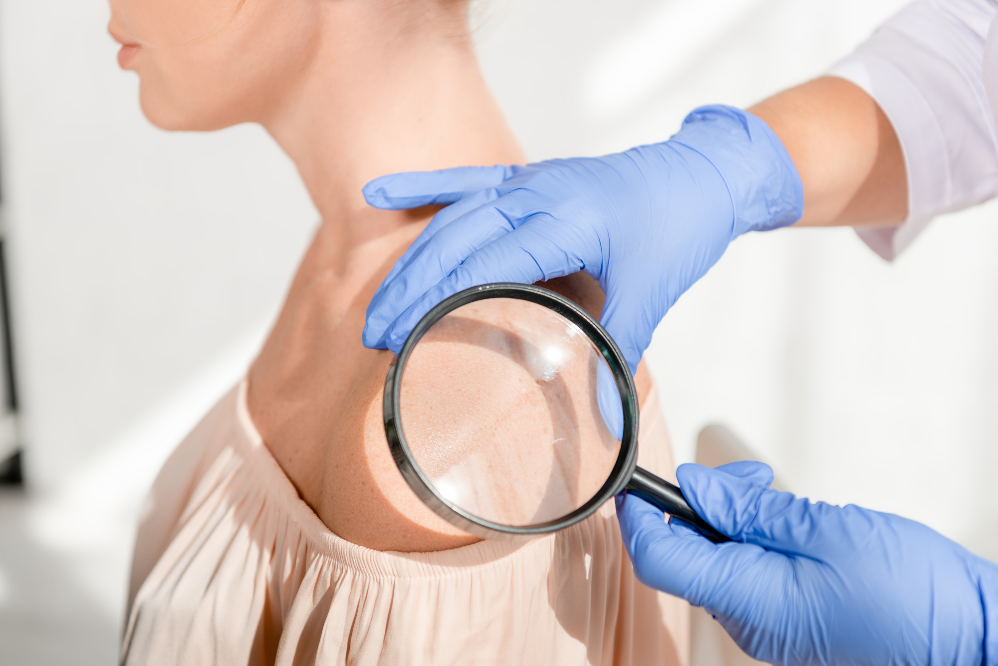
What is a Skin Growth?
The term skin growths encompass the lumps and bumps nearly everyone finds on their skin. In fact, the reason most patients make an appointment with the dermatologist is so we can “check this spot.” Lumps and bumps are the backbone of dermatology.
While the vast majority of the skin growths are benign (not cancerous), some are malignant (cancerous). The art of dermatology is being able to tell the difference in each type of growth and treat it accordingly.
The diagnosis of a skin growth can range from benign, pre-malignant, or malignant. Depending on this diagnosis, a necessary treatment course may require no intervention, intermediate intervention, or aggressive treatment.
What Causes Benign Skin Growths?
Skin growths are normal tissues that become overgrown. It’s a natural, evolutionary process. Babies have perfect, smooth, clear skin, but as we age, our skin changes. It can change shape, color, and texture. Sometimes, these changes are triggered by specific stimuli including sun exposure and viruses. Other times, skin changes simply due to age.
What Causes Malignant Skin Growths?
Malignant skin growths include a range of precancerous or cancerous growths that require treatment before they develop into a more complicated problem. Due to sun exposure, genetics, viral infection, or a combination of these factors, the cells have become abnormal. If left untreated, the cells will continue to mutate and move deeper into the skin.
What Are the Symptoms of Skin Growths?
Skin growths vary drastically in appearance based on the reason for the growth. They cover a huge spectrum of depth and severity:
- The depth can be superficial, intermediate, or extreme.
- The affected area can be a single spot or multiple spots.
Benign skin growths include:
Seborrheic Keratosis
The most common skin growth dermatologists see, this a classic case of overgrown skin. The external skin has overgrown and thickened as the person ages.
Cherry Angioma
These growths are uncommon in youth, but begin to appear on nearly everyone in their 20s and 30s (and older). Cherry angiomas show up as tiny, red capillary growths on faces, backs, forearms, and legs. If you look closely, you’ll probably find one some place on your body.
Warts
While seborrheic keratosis and angiomas are naturally occurring skin growths, warts are a virally induced skin growth. Warts appear due to exposure to an external trigger — the human papilloma virus (HPV). When any of the 100+ HPV subtypes contact the skin, the virus triggers an overgrowth of external skin. Because it’s external, the immune system doesn’t automatically respond to fight the virus and growth continues. The dermatologist chooses a treatment (such as freezing) to wake up the body, so it recognizes the virus and starts to fight it.
Cysts
A cyst is a ball of skin cells trapped under the skin. If these are non-symptomatic and not problematic, no treatment is necessary. However, if they become inflamed, tender, or occur in a problematic location, we remove them.
Lipomas
These non-cancerous growths are collections of thicker and compact fat that make bumps under the skin. Patients often present with multiple lesions.
Moles
Moles are clumps of pigmented cells that vary drastically in appearance and severity.
Some moles are congenital — children are born with the spot, or the spot appears within the first year of life. As the baby develops in utero, pigmented cells cover the baby — like a paint job. As these pigmented cells migrate over the skin, some cells clump up. These clumps are sometimes immediately recognized as congenital moles. However, when a mole activates later in life, it’s considered an “acquired” mole.
Mole treatment depends on the situation and diagnosis. Some moles can be left alone. Problem-causing moles, perhaps in a location where they are rubbed and aggravated, need to be removed. Other moles need to be biopsied and removed because of the potential danger it poses for developing into a cancerous cell.
Malignant skin growths include:
Precancerous Skin Growths
Precancerous skin growths are not yet dangerous. However, with either viral or skin exposure, the DNA in those cells starts turning abnormal. It then starts growing abnormally — developing texture, color, and sensitivity. These initially superficial growths can then push into the skin, which moves the classification from precancerous growth to in situ carcinoma, and eventually invasive skin cancer.
When we treat precancerous superficial growths aggressively, we’re able to prevent much deeper damage.
Basal Cell Carcinoma
Basal cells are typically located in sun-exposed areas and are triggered by sun exposure. While there are more basal cell carcinomas diagnosed per year than all other cancers combined, it’s also the easiest to treat. These classic pearl papules appear as clear bumps and can go undetected for months or years. When the skin finally starts breaking down, most people are prompted to visit their dermatologist.
Dermatologists can generally identify these as basal cell immediately. They will then conduct a biopsy to identify the subtype and decide on a reasonable next step for removing the growth.
Squamous Cell Carcinoma
Squamous cell carcinoma is triggered by UV rays and/or the human papilloma virus. HPV has a strong cancer-causing effect.
Squamous cell carcinoma often begins as precancerous actinic keratosis. If left untreated, it can grow into the epidermis and develops into in situ squamous cell carcinoma. If still untreated, it can become invasive.
We classify these progressions with three levels of squamous cell carcinoma:
- Well-differentiated: Cells look similar to normal skin but are atypical.
- Moderately differentiated: Cells start to lose the appearance of skin and break into more primitive looking tissue
- Poorly differentiated: The cells become so atypical, it starts to look like other tissue types. It can then spread through skin, lymphatics, or nerves.
Squamous cell carcinoma is primarily treated with surgery. Radiation and other techniques are occasionally used, but the vast majority are surgically removed.
Melanoma
Melanoma is known to be dangerous. While it’s not the worst cancer, it’s the most common dangerous cancer we see in dermatology.
Why is it so dangerous? It can be small, overlooked, and fatal if untreated. Yet dermatologists have become increasingly fast and precise at detecting, diagnosing, and treating melanoma. In fact, 9 out of 10 cases are detected superficially — that means they’re detected so early they have not yet penetrated the skin. At this point, its risk of developing into a harmful lesion is minimal.
The difficulty lies in detecting melanoma with varied color — they can appear pink, clear, or brown. In fact, some spots can look identical to actinic keratoses. As a precaution, dermatologists act very aggressively to leave no potential melanoma lesion untested and therefore untreated.
What Are The Treatment Options For Skin Growths?
The necessary treatment for skin growths depends on the type of growth and the discomfort or risk it poses. Not every skin growth needs to be removed. However, if we need to remove a growth for cosmetic or medical reasons, we select one of these treatments, depending on what’s most effective for the specific growth.
ED & C
Electrodesiccation and curettage, also known as ED & C, is a method for treating non-melanoma skin cancers through a serious of scraping and cauterizing techniques. When we identify basal and squamous cell carcinomas early, we often choose to treat them with ED & C. With this inexpensive and effective method, superficial, non-melanoma skin cancers can be easily removed.
Topical Chemotherapy
We opt to remove some growths with topical chemotherapy creams such as Imiquimod, Fluorouracil, and ingenol mubutate. These creams target abnormal cells and cause the skin cells to slough off. The prescribed dosage period is determined by the type of medication selected and how aggressively we need to treat the growth.
Photodynamic Therapy
Photodynamic therapy (PDT) uses light to treat skin growths. We apply a photosensitizing agent to the skin and allow it to absorb into the areas we want to remove. We then expose those areas to specific wavelengths of light that consequently destroy the targeted cells. The skin heals completely within 10 days or less.
Mohs Surgery
Mohs surgery is the standard of care for removing many types of skin cancer. In this outpatient procedure, we remove the visible growth and a thin peripheral margin of skin surrounding it. The surrounding tissue is examined to see if any further affected areas need to be removed. The procedure is repeated as necessary to remove all irregular borders of the suspicious growth. Depending on the location of the surgery, most patients heal within 2 weeks.
Radiotherapy
Patients who are poor surgical candidates may opt for radiotherapy to treat basal cell or squamous cell carcinoma. These abnormal cells are radiosensitive and often respond well to a 3-6 week course of radiotherapy.
Cryotherapy
With benign growths such as warts, angiomas, and precancerous lesions, cryotherapy is a viable treatment option. With this treatment, we apply liquid nitrogen that is so cold it destroys the cells. This procedure can be quite painful, but stinging and burning will subside after about 10 minutes. Damaged cells will slough off and heal within two weeks.
Oral Chemotherapy
While rarely used, some oral chemotherapy medications have been found effective in treating skin cancers. Your dermatologist will provide these options if necessary for the treatment of your skin growth.
FAQ’s About Skin Growths
Can benign growths turn into cancer?
Any cell in the body can turn into cancer. In dermatology, we work to identify precancerous and suspicious growths before they mutate into a problem that’s more difficult to treat.
I have atypical moles. Should I be concerned about skin cancer?
While more than 99% of moles are considered benign, like any cell in the body, they can turn cancerous. When benign moles turn cancerous, we call it melanoma. However, there’s a spectrum between fully benign moles and cancerous melanoma.
Many patients have slightly atypical moles that fall somewhere in between. These are not cancerous. They may not even be precancerous, but they are a marker for the potential of a cancer developing in the patient. If you have an atypical mole, see your dermatologist for a skin check.
My mole is benign, but I want it removed. Can I?
Yes — some are uncomfortable or get agitated and bleed. In these cases, we can remove the mole.
How can I spot melanoma?
Melanoma is best identified by a trained dermatologist. It presents with a variety of colors and appearances, making it difficult to distinguish from other types of growths. If your dermatologist suspects melanoma, they will biopsy the area and aggressively work to treat the growth according to the results.
Click here to find an Epiphany provider near you.

Dr. R. Todd Plott is a board-certified dermatologist in Coppell, Keller, and Saginaw, TX. His specialization and professional interests include treating patients suffering with acne, identifying and solving complex skin conditions such as psoriasis, rosacea, atopic dermatitis, and identifying and treating all types of skin cancers. In his spare time, Dr. Plott enjoys cycling, traveling with his wife, and spending time with his children and new grandson.
Learn more about Dr. Plott.